
Key Takeaways
-
Orthodontic X-rays reveal what a visual exam cannot, including tooth roots, unerupted teeth, impacted or missing teeth, jaw relationships, and bone support that guide safe treatment planning.
-
A panoramic X-ray shows all teeth and both jaws in one image and is often the first scan, while a cephalometric side-profile X-ray helps assess bite severity and jaw alignment.
-
Bitewing X-rays mainly check for cavities and cannot show tooth eruption or jaw relationships, so cavities should be treated before braces or aligners begin.
-
CBCT 3D scans are reserved for complex cases like difficult impactions and should not be used routinely when a lower-dose image can answer the clinical question.
-
Orthodontists follow the ALARA principle, using the fewest images and smallest area needed, and X-rays are taken based on clinical need rather than a fixed schedule.
-
Bringing recent dental X-rays from your general dentist can reduce repeat imaging, though orthodontists may still need panoramic or cephalometric views for jaw and bite details.
Ever wonder why your orthodontist wants a picture of what’s going on beneath your gums? You’re not alone! A smile may look perfectly clear from the outside, but a lot happens below the surface. Teeth have long roots, new teeth wait in the jaw, and the bones holding it all together are hidden from view.
That’s exactly why what orthodontic X-rays can help evaluate is such a popular question for parents, teens, and adults alike. These images give your orthodontist a clear look at things a simple exam can’t show. They help plan safe, smart, and personalized care.
In this friendly guide, we’ll walk through each type of X-ray and what it shows. We’ll also talk about safety, how often you might need images, and what to expect at your visit. By the end, you’ll feel confident and ready to ask great questions about your own treatment.
Why Orthodontists Use X-Rays in the First Place
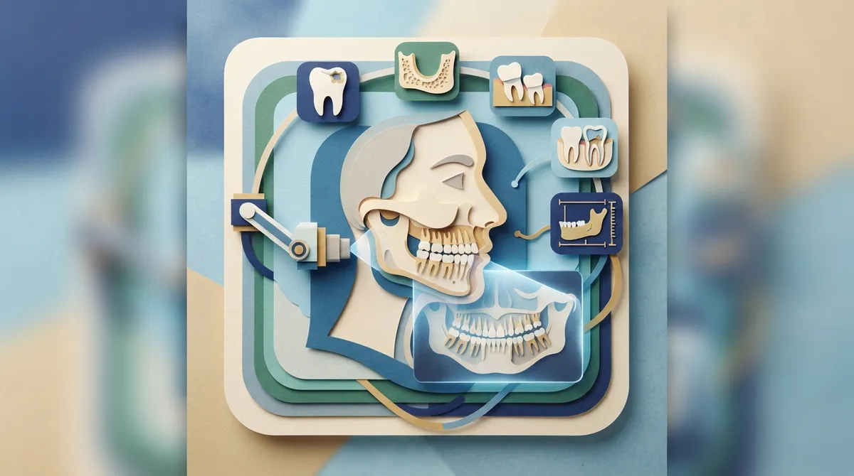
Think of an orthodontic exam like looking at a house from the street. You can see the paint and the windows. But you can’t see the foundation, the pipes, or the wiring. X-rays are how your orthodontist checks the “foundation” of your smile.
Orthodontic X-rays help assess structures that can’t be fully judged by eye alone. These include tooth roots, teeth that haven’t come in yet, the jawbones, and how the teeth relate to the bones of the face.
Here’s the good news. Imaging is not taken just for the sake of it. Each X-ray should be chosen for your specific needs and the questions your orthodontist needs answered. The goal is always to get the most helpful information with the least exposure.
What Orthodontic X-Rays Can Help Evaluate
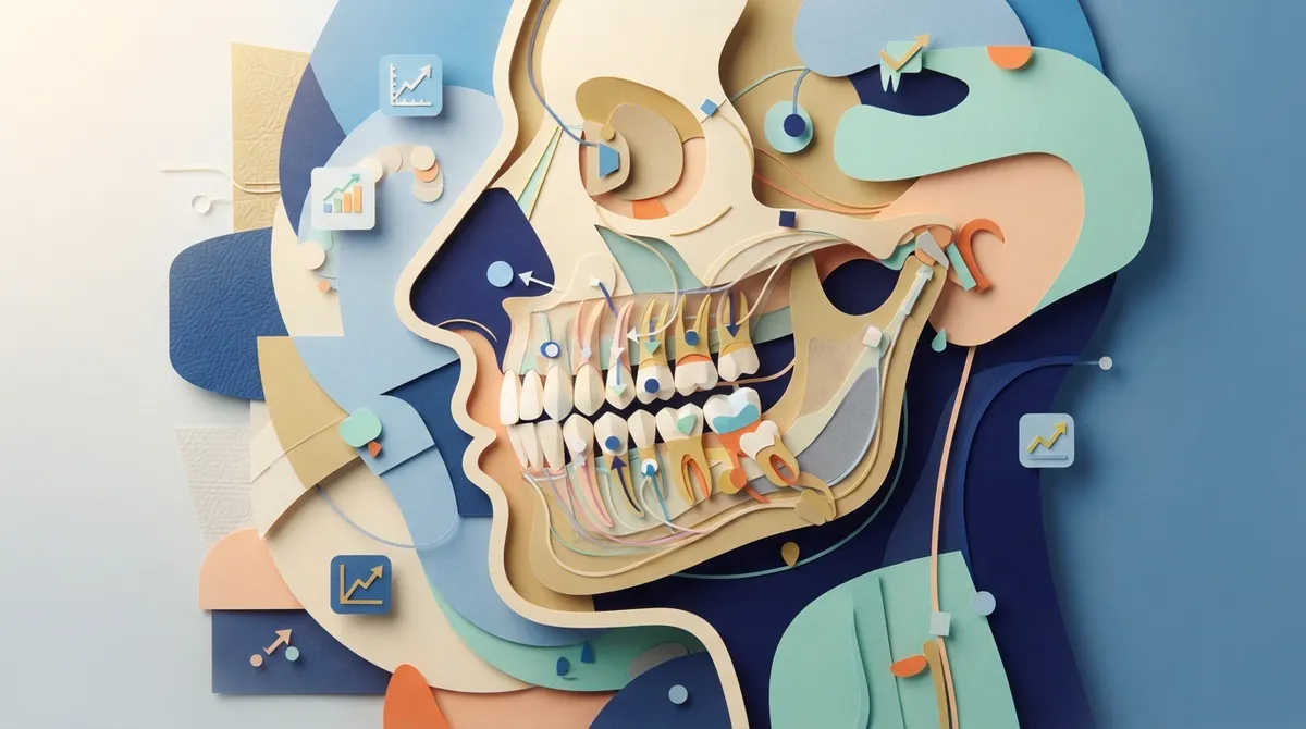
So, what can these images actually reveal? Quite a lot! Here are the main things your orthodontist can check with the right X-ray.
- Tooth development and eruption: Are baby teeth falling out on time? Are adult teeth growing in the right spots?
- Impacted teeth: Some teeth get stuck under the gums and can’t come in on their own.
- Missing or extra teeth: Sometimes a tooth never forms, and sometimes there’s an extra one hiding.
- Tooth roots: Root length, shape, and position all matter when moving teeth safely.
- Jaw relationships: X-rays show how the upper and lower jaws line up with each other.
- Bone support: Healthy bone around the teeth is key to a stable result.
- Growth patterns: For kids and teens, images help show how the face and jaws are developing.
These details guide the entire treatment plan. They help your orthodontist decide whether braces or Spark™ Clear Aligners are the better fit for your smile.
The Main Types of Orthodontic X-Rays
Not every X-ray does the same job. Each type answers a different question. Let’s break them down in plain language.
Panoramic X-Rays: The Big Picture
A panoramic X-ray is like a wide-angle photo of your whole mouth. The machine slowly moves around your head and captures all your teeth and both jaws in a single image.
This is often the first image taken at an orthodontic visit. It helps your orthodontist evaluate:
- Tooth development and eruption
- Impacted or unusually positioned teeth
- Missing or extra teeth
- Some jaw or dental abnormalities
For a parent of a 7-year-old, this one image can show if adult teeth are on track. That’s why it plays such a big role in early treatment decisions.
Cephalometric X-Rays: The Side Profile
A lateral cephalometric X-ray (often called a “ceph”) shows a side view of your head. It captures the skull, jaws, and teeth in profile, almost like a silhouette.
Orthodontists use it to look at:
- How the upper and lower jaws line up
- Skeletal patterns of the face
- The severity of a bite problem
- Craniofacial growth over time
This image is a favorite for planning treatment. For example, it can help show why someone has a deep overbite or an underbite. If you’re curious about bite corrections, our guide on how braces fix a deep overbite goes into more detail.
Periapical X-Rays: Up Close and Personal
A periapical X-ray zooms in on just a few teeth, showing each one from the crown all the way down to the root tip. It also shows the bone around the root.
Your orthodontist may request one when there’s a specific concern. It’s helpful for checking:
- Root shape and length
- Signs of root resorption (when a root starts to shorten)
- Disease or trouble around the root of a tooth
Think of it as a magnifying glass for one trouble spot.
Bitewing X-Rays: Checking the Back Teeth
Bitewing X-rays show the crowns of your upper and lower back teeth, plus the spaces between them. You bite down on a small tab, and that’s it!
They’re mainly used to check for cavities, and they can help assess bone levels. But here’s an important point: bitewings are not a substitute for broader orthodontic imaging. They can’t show tooth eruption or jaw relationships the way a panoramic or ceph image can.
That’s one reason healthy teeth matter before treatment starts. Cavities should be handled before braces or aligners begin.
CBCT: The 3D View
Cone-beam computed tomography, or CBCT, creates a three-dimensional picture of your teeth, bones, and jaws. It’s a powerful tool, but it’s not for everyone.
It may be considered for special situations, such as:
- Complex impacted teeth
- Anatomy that regular images can’t show clearly
CBCT should not be used routinely when a lower-dose image can answer the question. In other words, it’s reserved for when it truly adds value.
Quick Comparison of Orthodontic X-Ray Types
| X-Ray Type | What It Shows | Best For |
|---|---|---|
| Panoramic | All teeth and both jaws in one view | Tooth development, impactions, missing or extra teeth |
| Cephalometric | Side profile of skull, jaws, and teeth | Jaw relationships, bite severity, growth |
| Periapical | Individual teeth from crown to root | Root shape, resorption, root-area concerns |
| Bitewing | Crowns of back teeth and spaces between | Cavities and bone levels |
| CBCT (3D) | Three-dimensional view of teeth and bone | Complex impactions and hard-to-see anatomy |
Are Orthodontic X-Rays Safe?
This is one of the most common questions we hear, especially from parents. The short answer is yes, when used wisely. Orthodontists follow a guideline called ALARA, which stands for “as low as reasonably achievable.” That means using the fewest images and the smallest area needed to get useful information.
Here’s some helpful context. According to the British Orthodontic Society’s 2016 guidance, approximate effective-dose ranges are:
- Panoramic X-rays: about 0.0027 to 0.038 mSv
- Lateral cephalometric X-rays: about 0.0022 to 0.0056 mSv
- Periapical or bitewing X-rays: about 0.0003 to 0.022 mSv
Actual dose varies by equipment and technique. The U.S. FDA also notes that dental X-rays account for roughly 2.5% of the effective dose from medical X-rays and fluoroscopies. It recommends taking only the images needed for diagnosis.
The American Dental Association also supports individualized decisions about imaging. In plain terms, you shouldn’t get X-rays automatically on a fixed schedule. They should be based on your needs.
What to Expect During Your Orthodontic X-Ray Visit
Good news: getting X-rays is quick and painless. Most people finish in just a few minutes. Here’s how it usually goes.
- Your orthodontist reviews your needs. They decide which images will answer your questions.
- You get comfortable. You may wear a protective apron and remove glasses or earrings.
- The image is taken. For a panoramic or ceph, you stand still for several seconds while the machine moves around you.
- The images appear on screen. Digital images are ready almost right away.
- You talk through the results. Your orthodontist explains what they see in simple terms.
At our office, we use modern digital technology. That means quick images, clear results, and a comfortable experience for the whole family. Want a preview of the process? Check out what to expect at your first visit.
How X-Rays Help Different Patients
Every smile has its own story, and X-rays help tell it. Here’s how they help different people.
For Kids and Younger Children
X-rays can spot problems early, like crowding, impacted teeth, or jaw growth concerns. Catching these early may make later treatment simpler. This is a big part of orthodontics for kids.
For Teens
Teens often have most of their adult teeth in. Images confirm everything is in place and help plan the right type of adolescent treatment, whether that’s braces or clear aligners.
For Adults
Adults may have fillings, crowns, or gum concerns. X-rays help check bone health and root condition before moving teeth. This makes adult treatment safer and more predictable.
For Patients Needing Retreatment
If you had braces years ago and your teeth have shifted, images can show what changed. They help your orthodontist build a fresh plan that fits you now. And a good retainer routine can help protect your results, so retainers are worth discussing too.
How Often Do You Need X-Rays During Treatment?
Here’s something that surprises many people: you don’t need X-rays at every appointment. Images should be taken based on clinical need, not a rigid calendar.
Most patients have a set of images at the start to build a plan. Additional images may be taken if something specific needs a closer look, such as checking root health or tooth movement. Many patients also have a final set near the end of treatment.
If you already have recent dental X-rays from your general dentist, bring them along! Your orthodontist may be able to use them, though they may still need other views for jaw and bite details.
Tips to Get the Most Out of Your X-Ray Appointment
- Bring past images: Recent dental X-rays can sometimes reduce repeat imaging.
- Share your health history: Tell us about any health changes or if you may be pregnant.
- Ask questions: Wondering why a certain image is needed? Just ask!
- Hold still: Staying steady helps get a clear image the first time.
- Remove metal items: Jewelry and glasses can get in the way.
Your Smile Starts With a Clear Picture
Orthodontic X-rays are a helpful, low-dose way to see what’s hiding beneath the surface. They help your orthodontist check tooth roots, unerupted teeth, jaw relationships, and so much more. With the right image for the right reason, your care plan can be safer and more personal.
At Brockway Orthodontics, Dr. Brockway takes the time to explain every step in a friendly, easy-to-understand way. You’ll always know why an image is needed and what it shows. If you’d like to see how it all works, you can visit us on Google — Brockway Orthodontics to see what our patients are saying. You can also learn more about Dr. Brockway and our services.
Ready to take the first step toward a confident smile? Book your appointment online today, or contact us with any questions. We can’t wait to meet you!
FAQs
Q: What can an orthodontic X-ray show?
A: Orthodontic X-rays can show tooth roots, teeth that haven’t come in yet, impacted or missing teeth, jaw relationships, and bone support. They reveal things your orthodontist can’t see during a regular exam. That helps us plan safe, personalized care.
Q: What is the difference between a panoramic and a cephalometric X-ray?
A: A panoramic X-ray gives a wide view of all your teeth and both jaws at once, which is great for checking tooth development and eruption. A cephalometric X-ray shows a side profile of your skull, jaws, and teeth. It helps with jaw relationships and bite severity.
Q: When is a CBCT scan needed for orthodontic treatment?
A: A CBCT scan is a 3D image that’s saved for special cases, like complex impacted teeth or anatomy that regular images can’t show well. It isn’t used routinely. If a lower-dose image can answer the question, that’s the better choice.
Q: Are dental X-rays safe for children and teens?
A: Yes, when used wisely. Orthodontists follow the ALARA principle, using the fewest images and smallest area needed for a clear diagnosis. Doses from orthodontic X-rays are low, and images are only taken when they’re truly helpful.
Q: Do I need new orthodontic X-rays if I already have recent dental X-rays?
A: Not always! Bring your recent images, and your orthodontist can review them. You may still need other views, like a panoramic or cephalometric image, to see jaw and bite details that regular dental X-rays don’t cover.
Our Service Area