Key Takeaways
-
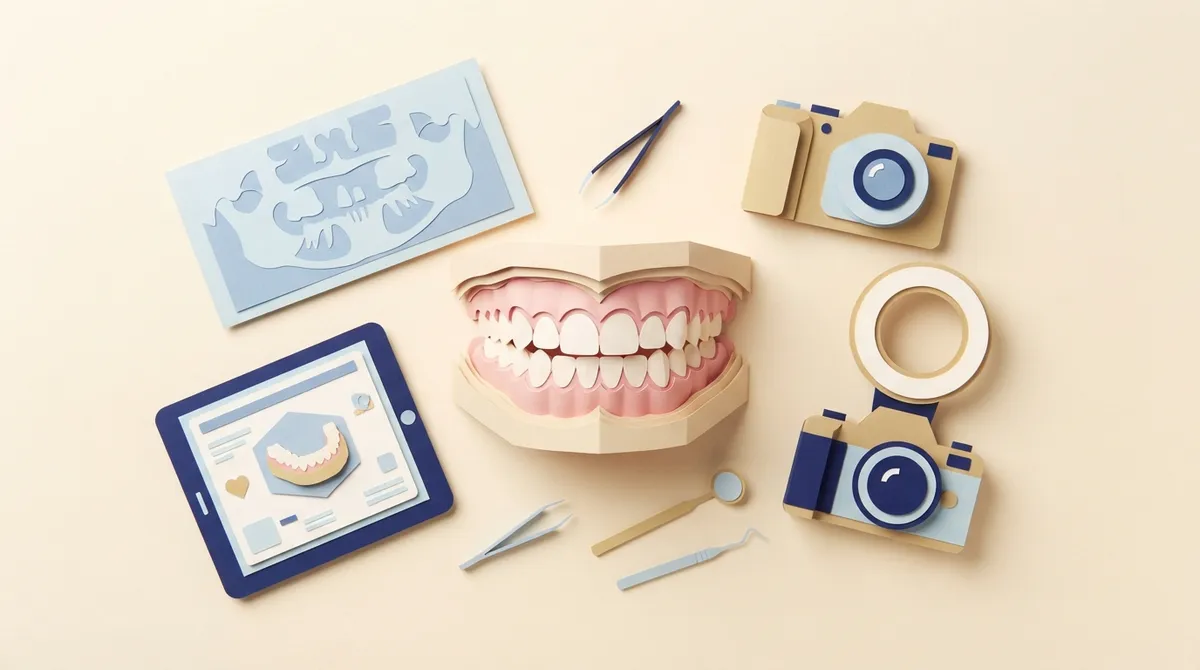
A thorough clinical exam and health history is the foundation of assessing a developing bite, covering jaw relationships, crowding, overbite, crossbite, and which teeth have erupted.
-
Records are not a fixed package; the mix depends on the child's age, history, and exam findings, so no child needs every image type.
-
Extraoral and intraoral photos, plus digital scans or models, create a baseline that lets orthodontists track changes as the child grows.
-
A panoramic X-ray is often the most useful radiograph for a developing bite because it shows unerupted, missing, or extra teeth, but X-rays should be taken only when clinically justified.
-
Cephalometric X-rays and CBCT 3D scans are reserved for specific situations, such as tracking jaw growth or evaluating an impacted tooth, and are not routine.
-
The American Association of Orthodontists recommends a check-up by about age seven, and signs like early baby tooth loss or chewing trouble warrant an earlier evaluation.
If your child has a wiggly tooth, a crowded smile, or a bite that just looks a little “off,” you may wonder what an orthodontist actually looks at. It’s a great question! The good news is that checking a growing bite is not a one-size-fits-all process. It starts with a friendly conversation and a careful look, and then adds a few helpful records to build the full picture.
So, what records help assess a child’s developing bite? The short answer: a clinical exam and health history, photographs, dental models or digital scans, and, only when needed, X-rays. Each record tells a different part of the story. Together, they show how your child’s teeth, jaws, and face are growing.
In this guide, we’ll walk through each record in plain language. You’ll learn what it shows, why it matters, and when it’s used. Whether you are a parent of a seven-year-old or a teen who is curious about your own bite, you’ll know exactly what to expect. At Brockway Orthodontics, we love when families feel informed and relaxed, so let’s dive in!
Why Records Matter for a Growing Smile
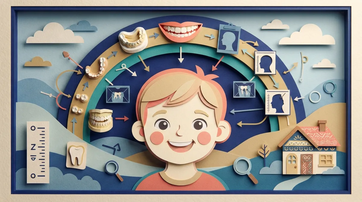
A child’s mouth is always changing. Baby teeth fall out, adult teeth come in, and the jaws keep growing. Because of this, one quick glance isn’t enough to understand what’s going on.
Records give your orthodontist a clear snapshot of today. They also create a baseline, which means a starting point to compare against later. When we look at your child again in six months or a year, we can see what has changed and what hasn’t.
The American Association of Pediatric Dentistry (AAPD) describes three broad areas of orthodontic evaluation in the developing dentition:
- Health of the teeth and oral structures (gums, tissues, and tooth condition)
- Tooth alignment and occlusal relationships (how the teeth line up and meet)
- Facial and jaw proportions (how the jaws and face fit together)
Every record we collect helps answer questions in one or more of these areas. And here’s something that surprises many parents: there is no fixed “package” of images every child must have. The records chosen depend on your child’s history, age, and what we see during the exam.
Step One: The Clinical Exam and Health History
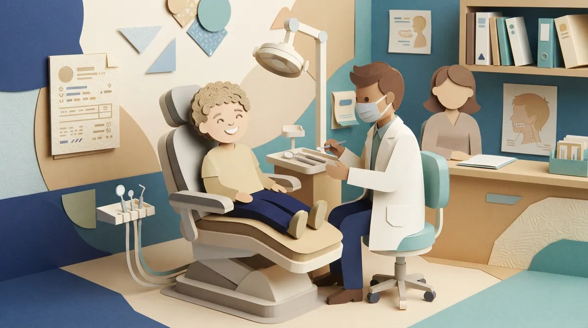
Before any pictures or scans, the most important record is the exam itself. This is where your orthodontist listens, looks, and gently checks how your child’s mouth works.
We usually start by asking about your child’s health, past dental care, habits like thumb sucking, and any trouble with chewing, speech, or jaw comfort. You know your child best, so your observations are truly valuable here.
What We Look for During the Exam
During a clinical exam, the orthodontist may check:
- Facial symmetry and side profile
- How the upper and lower jaws relate to each other
- Whether the center lines of the teeth match up
- Crowding or extra spacing between teeth
- Overbite and overjet (how far the top teeth cover or stick out past the bottom)
- Crossbite or open bite patterns
- Which teeth have come in and which are still on their way
- Gums and other oral tissues
- Chewing, swallowing, and jaw movement
This hands-on look is the foundation of everything else. In many cases, the exam tells us a lot before we ever need a single image. If you’d like to know what a first appointment feels like, our Your First Visit page explains the experience step by step.
Photographs: A Picture Really Is Worth a Lot
Photographs are one of the simplest and most useful records. They’re quick, comfortable, and give us a lasting visual reference.
Orthodontists usually take two kinds of photos:
- Extraoral photos show your child’s face from the front and side. These help us look at facial balance, smile shape, and profile.
- Intraoral photos show the teeth and bite from different angles, such as front, left, right, and the top and bottom arches.
These pictures supplement the exam rather than replace it. They let us see details we can study later, and they make it easy to compare your child’s progress over time. Many parents love looking back at these “before” photos once treatment is done!
Dental Models and Digital Scans
Next up are records that show the shape of your child’s teeth and how the upper and lower arches fit together.
In the past, this meant biting into trays of goopy material to make plaster models. Today, many practices use digital intraoral scans instead. A small wand gently captures the teeth, and a 3D picture appears on screen. It’s quick, comfortable, and no messy molds are needed. At Brockway Orthodontics, we’re big fans of this modern approach because kids tend to find it far more fun than traditional impressions.
What Models and Scans Can Show
Whether made from plaster or captured digitally, these records help us assess:
- How the upper and lower teeth meet when your child bites down
- Arch-length concerns, meaning whether there is enough room for all the teeth
- Tooth-size differences between the upper and lower teeth
- Arch asymmetry, where one side looks different from the other
Because a 3D model can be rotated and studied from every angle, it helps with careful planning. It also gives us another baseline to compare against as your child grows.
X-Rays: Helpful When Needed, Not Automatic
X-rays are a hot topic for many parents, so let’s be clear. Radiographs (the technical word for X-rays) are chosen only when they are clinically indicated. That means we use them when they will truly help us make a better diagnosis.
The AAPD recommends that the timing and frequency of X-rays be based on each child’s individual history, clinical findings, and diagnostic needs. There is no universal number of images that every child must have. The goal is to take the minimum needed for good care.
Panoramic X-Rays
A panoramic X-ray is a single wide image that shows all the teeth, both jaws, and the areas where adult teeth are still developing under the gums. It can help us see:
- Stage of dental development and eruption
- Teeth that haven’t come in yet
- Missing or extra teeth
- Unusual tooth shapes or positions
- Signs of other concerns that need attention
This is often the most helpful X-ray for a child with a developing bite, since it shows what is happening below the gumline.
Intraoral X-Rays
These are smaller images that focus on specific teeth. They give a closer look at individual areas when we need extra detail about development, eruption, or tooth health.
Cephalometric X-Rays
A cephalometric (or “ceph”) X-ray is a side-view image of the head. It helps us study how the teeth and jaws relate to each other in different dimensions. We may use one when we need to understand jaw growth patterns and set a baseline for tracking growth. It’s not needed for every child, only when the information will guide our decisions.
What About 3D Scans (CBCT)?
CBCT is a 3D imaging option. It is not a routine screening record. We may consider it for specific problems, such as a tooth that is stuck below the gum (an impacted tooth), and only when the added information is truly justified.
Quick Comparison: Which Record Shows What?
Here’s a handy table to keep all of this straight.
| Record | What It Helps Show | Used for Every Child? |
|---|---|---|
| Clinical exam and history | Overall oral health, bite, jaw and face balance, growth stage | Yes, this is the starting point |
| Extraoral and intraoral photos | Facial appearance, teeth, and bite from several views; baseline for later comparison | Commonly used as a supplement |
| Dental models or digital scans | Arch shape, bite relationship, crowding, arch asymmetry | Depends on findings |
| Panoramic or intraoral X-rays | Tooth development, eruption, missing or extra teeth, tooth anomalies | Only when clinically justified |
| Cephalometric X-ray | Jaw and tooth relationships, growth baseline | Only when needed |
| CBCT (3D imaging) | Detailed view of specific problems, like an impacted tooth | No, not a routine record |
How the Records Work Together
No single record tells the whole story. A photo can show how a smile looks, but not what’s under the gums. An X-ray can show hidden teeth, but not how your child chews. That’s why orthodontists read all the records together.
Think of it like a puzzle. The exam gives us the corner pieces, photos and scans fill in the edges, and X-rays add the middle pieces only when we need them. Once the picture is clear, we can explain what we see in simple terms and talk about whether to watch, wait, or begin early treatment.
Records also aren’t a one-and-done event. Because a child’s mouth keeps changing, we revisit and update them as the dentition develops. A bite that looks fine at age seven may need a closer look at age nine or ten. Regular check-ins keep us a step ahead.
When Should a Child Be Evaluated?
Many families ask when to schedule that first visit. The American Association of Orthodontists recommends a check-up by around age seven, which is when a mix of baby and adult teeth gives us helpful clues about how the bite is forming. That said, every child is different, and some families come in earlier because of a specific concern.
Signs that it may be time for an evaluation include:
- Early or late loss of baby teeth
- Difficulty chewing or biting
- Mouth breathing or thumb sucking that continues
- Teeth that look crowded or stick out
- Upper and lower teeth that don’t meet properly
- Jaws that shift or make sounds
Coming in doesn’t mean your child needs braces right away. Often, the best answer is simply to monitor growth. For more guidance, our article on how to know when your child should first see an orthodontist is a helpful read, and our Orthodontics for Kids guide covers what every parent should know.
Tips to Make Record-Taking Easy for Your Child
Worried your child might feel nervous? That’s completely normal, and a little preparation goes a long way. Try these ideas:
- Explain it simply. Tell your child we’ll take some pictures and look at their teeth. Skip scary words.
- Bring a list of questions. Jot down anything you’ve noticed, like grinding, snoring, or chewing trouble.
- Share health history. Bring a note about past dental work, allergies, or medications.
- Stay positive. Kids pick up on our feelings. A calm, cheerful attitude helps them relax.
- Ask us anything. We’re happy to explain why each record is being taken.
Our team at Brockway Orthodontics takes the time to walk families through every step, and Dr. Brockway makes sure both parents and kids feel comfortable and informed. You can learn more about her approach on the Meet Dr. Brockway page.
The Role of Modern Technology
Today’s tools make collecting records easier than ever. Digital scans replace messy impressions, and 3D imaging helps with precise planning when it’s truly needed. For kids, this often means a faster, more comfortable visit.
Every piece of technology serves one purpose: helping us understand your child’s bite so we can recommend the right care at the right time. We always aim to use only what is necessary, especially when it comes to X-rays. You can see more about how a visit works in our What to Expect During Your Orthodontic Exam article.
If you’d like to understand how oral health organizations view dental care in general, the American Dental Association offers helpful public resources for families. And for professional standards in our field, the Florida Association of Orthodontists is a great place to learn more.
Why Choose Brockway Orthodontics for Your Child’s Bite Evaluation?
When you’re looking for the right orthodontist in Clearwater, Florida, you want someone who is skilled, gentle, and honest. Dr. Brockway is all of those things. She truly listens to your concerns and takes time to explain what she sees, so you never feel rushed or confused.
We welcome the whole family, from kids with their first set of braces to adults looking for a discreet option. Our services include metal braces, ceramic (clear) braces, and Spark™ Clear Aligners, an affordable clear aligner choice for teens and adults. For children, we focus on careful monitoring and recommending treatment only when it truly helps.
Want to see what other families think? You can visit our Brockway Orthodontics location on Google and read reviews from our Clearwater community. And if you have questions about cost, our financing and insurance page explains the options available.
Conclusion: A Clear Picture Leads to Confident Care
So, what records help assess a child’s developing bite? A thorough clinical exam and health history come first, followed by photographs, dental models or digital scans, and X-rays only when they’re clinically justified. Cephalometric images and 3D scans are reserved for specific situations. Most importantly, these records are read together and updated as your child grows.
The best part? You don’t have to figure it out alone. A simple visit can answer your questions, ease your worries, and give you a plan for your child’s smile. Ready to take the first step? Book your child’s appointment online and let our friendly team at Brockway Orthodontics show you how easy it can be. You can also follow our smiles and updates on Facebook.
FAQs
Q: What does an orthodontist check at a child’s first bite evaluation?
A: We start with a gentle exam and health history. We look at the teeth, bite, jaw and facial balance, which teeth have come in, and how your child chews and moves their jaw. Photos or scans may be added, and X-rays only if they’re truly needed.
Q: Do children need X-rays to assess their developing bite?
A: Not always! X-rays are used only when they’re clinically justified, such as to check for missing, extra, or unerupted teeth. Many bite concerns can be understood from the exam, photos, and scans alone.
Q: What can a panoramic X-ray show about a child’s teeth?
A: A panoramic X-ray gives a wide view of all the teeth and both jaws, including adult teeth still under the gums. It helps us check development, eruption, and any missing or unusual teeth.
Q: How do photos and digital scans help assess a child’s bite?
A: Photos capture the face, teeth, and bite from several angles, while digital scans create a 3D model of the arches. Together they show how the teeth fit, help us spot crowding or asymmetry, and give us a baseline to compare as your child grows.
Q: How often should a child’s developing bite be monitored?
A: It depends on your child’s history and what we see at each visit. Because growth keeps changing the bite, we revisit records over time and schedule check-ins that fit your child’s needs.
Our Service Area