Key Takeaways
-
CBCT is a 3D X-ray that is not routine for most orthodontic patients; it is discussed only when a specific question cannot be answered by an exam and regular flat X-rays.
-
The key test is whether the 3D detail could change the diagnosis or treatment plan; if the scan would not change anything, it usually is not needed.
-
The most common reason is an impacted tooth, such as an upper canine, where 3D imaging shows its exact position and helps plan how to guide it into place.
-
A 2024 systematic review found adjacent-root resorption in 29.9% of impacted canines assessed with CBCT versus 15.2% with panoramic X-rays, showing why 3D detail matters when root damage is a concern.
-
CBCT involves more radiation than many standard dental X-rays, so patients should ask for the smallest field of view and lowest settings that still give a clear image.
-
Patients can ask what question the scan answers, whether existing X-rays were reviewed first, and how the result could change their plan.
If your orthodontist has ever said the words “3D scan,” you may have wondered whether it is a big deal. Good news: for most people, it is not. But there are times when a closer, three-dimensional look can really help. So, when might a CBCT scan be discussed in orthodontics? In short, it comes up when a specific question cannot be answered well with an exam and regular flat X-rays, and when the extra detail could change your plan.
CBCT stands for cone-beam computed tomography. It gives your orthodontist a 3D view of teeth, roots, and jaw structures. Think of it like the difference between a photo of a house and a full set of building plans. You do not need the plans to paint a wall. You might want them to move one.
In this guide, we will walk through how the decision is made, what situations call for a scan, and how to ask smart questions. Whether you are a parent, a teen, or an adult thinking about treatment in 2026, this will help you feel prepared and calm.
What Is a CBCT Scan, in Plain Words?
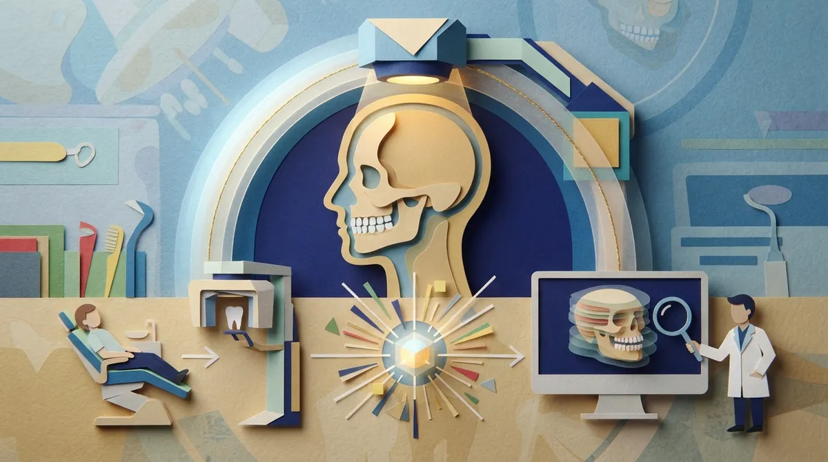
A CBCT scan is a special type of X-ray. A machine rotates around your head and takes many images in one pass. A computer then builds them into a 3D picture. Your orthodontist can look at it from any angle and scroll through it in thin slices.
A regular dental X-ray, like a panoramic image, squashes everything into one flat picture. Things can overlap or hide behind each other. CBCT removes much of that guesswork for a chosen area.
Why it is not an everyday scan
CBCT is not a routine record for every braces or aligner patient. Exams, photos, digital scans, and conventional X-rays are the usual starting point. Your care team should only suggest a CBCT scan when the benefit is expected to outweigh the added radiation. That is a careful, patient-first way to work.
The Simple Rule: Ask Whether the Answer Could Change Your Plan
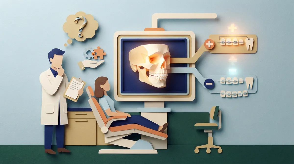
Here is the easiest way to think about it. An orthodontist may discuss a CBCT scan when three things are true:
- There is a specific clinical question that regular images cannot answer clearly.
- The 3D detail could change the diagnosis, the treatment plan, or how risks are managed.
- The scan can be limited to the area of interest, using the smallest field of view that works.
If the scan would not change anything, it usually is not needed. That is a fair question to ask at any visit. A thoughtful orthodontist will happily explain the reason.
When Might a CBCT Scan Be Discussed in Orthodontics? Common Situations
Let us look at the most common reasons. Remember, these are case-specific examples, not a checklist that applies to everyone.
1. An impacted or unerupted tooth
This is the classic example. An impacted tooth is one that is stuck under the gum and is not coming in on schedule. Upper canines, the pointed teeth beside the front teeth, are the most common ones. A flat X-ray may show that a tooth is missing from the smile. It may not show exactly where it is hiding or which way it is tilted.
With 3D detail, your orthodontist can see whether the tooth sits toward the cheek or the tongue side. That helps plan how to guide it down. It also helps when a dentist or oral surgeon needs to expose the tooth so a bracket can be attached.
2. Checking neighboring roots
Sometimes an impacted tooth presses on the root of the tooth next to it. This is called root resorption, and it means the root is slowly being worn away. It is not always easy to spot on a flat image.
A 2024 systematic review of impacted maxillary canines reported adjacent-root resorption in 29.9% of cases assessed with CBCT, compared with 15.2% using panoramic X-rays. That review looked at selected impacted-canine cases, so it does not mean this happens to most patients. It does show why 3D imaging can be useful when there is a real concern.
3. Suspected root problems or other findings
If an exam or standard image raises a question about a root, a cyst, or another finding, a limited CBCT may be discussed. For routine questions about root shape or tip-of-the-root health, small periapical X-rays are often the better first choice. CBCT is a next step only if those images do not give a clear answer.
4. Planning surgical exposure or traction
When a tooth needs to be pulled into place with gentle force, the exact position matters. A 3D view helps the team choose the safest path for the tooth. It can also help the surgeon plan a smaller, more precise procedure.
5. Selected complex jaw and facial cases
CBCT may also be considered for a few special situations, such as:
- Craniofacial differences or cleft-related care
- Noticeable facial or jaw asymmetry
- Planning for jaw (orthognathic) surgery
In these cases, the exact imaging depends on the clinical question. Your orthodontist will work with other specialists when needed.
When CBCT Is Usually Not the Starting Point
Most orthodontic patients never need one. For everyday crowding, spacing, or bite concerns, orthodontists generally rely on a careful exam, photos, a digital scan of your teeth, and standard X-rays. If you are curious about the basics, our guide on what orthodontic X-rays can help evaluate in 2026 breaks it down nicely.
A 2018 systematic review focused on children reached a helpful conclusion: CBCT is justified when regular X-rays cannot provide a correct diagnosis of the problem at hand. It does not support scanning every child before treatment. In other words, “because we have the machine” is never a good reason.
CBCT vs. Regular X-Rays: A Quick Comparison
This table shows how the common tools differ. Doses and details vary by machine and settings, so treat this as a general guide.
| Imaging type | What it shows well | Common orthodontic use | Limits |
|---|---|---|---|
| Panoramic X-ray | Wide, flat view of all teeth and jaws | Checking eruption and overall development | Overlapping structures, no true depth |
| Periapical X-ray | Close view of a few teeth and roots | Root shape, blunting, resorption, tip-of-root findings | Small area, flat image |
| CBCT (limited field) | 3D view of teeth, roots, bone | Impacted teeth, root questions, surgical planning | Higher dose than many dental X-rays, needs careful reading |
How to Prepare Smart Questions Before Your Visit
You do not need to be an expert to speak up. A few friendly questions can help you understand the plan and feel in control. Here is a simple way to approach it.
- Ask what question the scan is meant to answer. A clear reason shows the scan is purposeful.
- Ask whether existing images were reviewed first. Conventional X-rays and exams should usually come before 3D imaging.
- Ask how the result could change your plan. If the answer is “it might not,” that is worth discussing.
- Ask about the area being scanned. A smaller field of view means a more focused scan.
- Ask who will review the full image. A scan captures a volume of anatomy, so it should be read carefully.
If you are heading to a first appointment, our tips on how to prepare for your orthodontist consultation in Clearwater pair well with this list. For more ideas, see 13 questions to ask at an orthodontic consultation.
Understanding the Radiation Question
It is natural to ask about radiation. CBCT usually involves more radiation than many standard dental X-rays. The exact amount varies a lot by the machine, the settings, the size of the area, and the patient. That is why no single number fits every scan.
Good practice follows one idea: use the lowest exposure that still gives a clear, useful image. Your orthodontist should justify each scan one patient at a time. For children, the bar for choosing a CBCT is even more careful, since younger patients are more sensitive to radiation.
What this means for you
- A scan should have a clear purpose tied to your care.
- It should focus on the smallest area needed.
- It should be used only when simpler images are not enough.
You can learn more about general dental X-ray safety from the American Dental Association.
What Happens During a CBCT Scan?
Many people picture a scary tunnel, but a dental CBCT is much friendlier. Here is what a typical visit looks like:
- You stand or sit in the machine, and the team positions your head so it stays still.
- You may wear a lead apron or similar protection, depending on the office routine.
- The scanner rotates around your head once. This often takes under a minute.
- The computer builds the 3D picture, and your orthodontist reviews it with you.
It does not hurt, and there are no needles. The hardest part is holding still, which most kids and adults can manage fine.
Who Might Hear About CBCT? Examples for Different Families
Every person is different, so here are a few friendly examples.
- A parent of a 12-year-old notices that one canine has not come in. The orthodontist may first check with a panoramic image, then discuss a limited scan if the tooth looks tricky to guide.
- A teen with a baby tooth that will not fall out may need a closer look at the tooth underneath. You can read more about this in our post on retained baby teeth questions parents ask.
- An adult returning for retreatment may have a concern about a root or a previous issue. A scan is discussed only if standard images leave questions.
- A patient with a complex jaw concern might be a candidate for 3D imaging as part of a team plan with other specialists.
In most of these cases, the final decision comes after a careful exam. If you want to understand how that exam works, our article on what to expect during your orthodontic exam is a helpful read.
Why Reading the Scan Carefully Matters
A CBCT captures a whole volume of anatomy, not just the tooth in question. That means other findings can show up, like something in a sinus or near a joint. A responsible clinician reviews the full image, notes anything unusual, and arranges follow-up or a referral when it is needed. This is part of keeping you safe, and it is another reason scans should be done only when they matter.
How Modern Digital Tools Fit In
Many people mix up CBCT with other digital tools. They are different. A digital scanner takes a picture of the surface of your teeth with a small wand, no radiation involved. It replaces messy molds and helps plan braces and aligners. You can read more in our piece on Medit intraoral scanning for ortho. A CBCT, on the other hand, sees beneath the surface into roots and bone. Both are useful, and they answer different questions.
Your Care at Brockway Orthodontics
At Brockway Orthodontics in Clearwater, Dr. Brockway believes you should understand every step of your care. The practice uses modern technology, including digital impressions and 3D imaging, to plan treatment with precision and comfort. Imaging choices are made with your needs in mind, and your questions are always welcome. If you would like to see how others feel about their visits, you can visit our Brockway Orthodontics location on Google and read reviews.
The Florida Association of Orthodontists is also a good place to learn more about orthodontic care in our state.
Key Takeaways to Remember
- CBCT is a 3D scan, not a routine step for every patient.
- It is most often discussed for impacted teeth, root concerns, surgical planning, and selected complex cases.
- Exams and conventional X-rays usually come first.
- Each scan should be justified, kept as small as possible, and read carefully.
- You are always welcome to ask why a scan is suggested.
Ready to Talk It Through?
If you have questions about imaging, an impacted tooth, or your own treatment options, we are happy to chat. A friendly conversation is the best way to learn what fits your smile. Book your appointment online or contact our team to get started with Dr. Brockway in Clearwater, FL. We cannot wait to meet you.
FAQs
Q: Does every orthodontic patient need a CBCT scan?
A: Nope, not at all! Most patients never need one. Exams and regular X-rays are the usual starting point, and a CBCT is only considered when a specific question needs a 3D answer.
Q: Why might an orthodontist recommend CBCT for an impacted canine?
A: A flat X-ray cannot always show exactly where a hidden tooth sits or which way it is tilted. A limited 3D scan helps your orthodontist see its position, check nearby roots, and plan the safest way to guide it into place.
Q: Is CBCT safer or more accurate than a panoramic dental X-ray?
A: It is more detailed for certain questions, but it is not always safer, since the radiation dose is usually higher than many standard dental X-rays. That is why it is used only when the extra detail could change your care.
Q: How much radiation does an orthodontic CBCT scan involve?
A: There is no single number, because the dose depends on the machine, the settings, the size of the scanned area, and the patient. Your orthodontist should use the smallest field of view and lowest settings that still give a clear image.
Q: Is CBCT used routinely for braces or clear aligners?
A: No. Braces and clear aligners are usually planned with an exam, photos, a digital scan, and standard X-rays. A CBCT is saved for special cases like impacted teeth, root concerns, or surgical planning.
Our Service Area